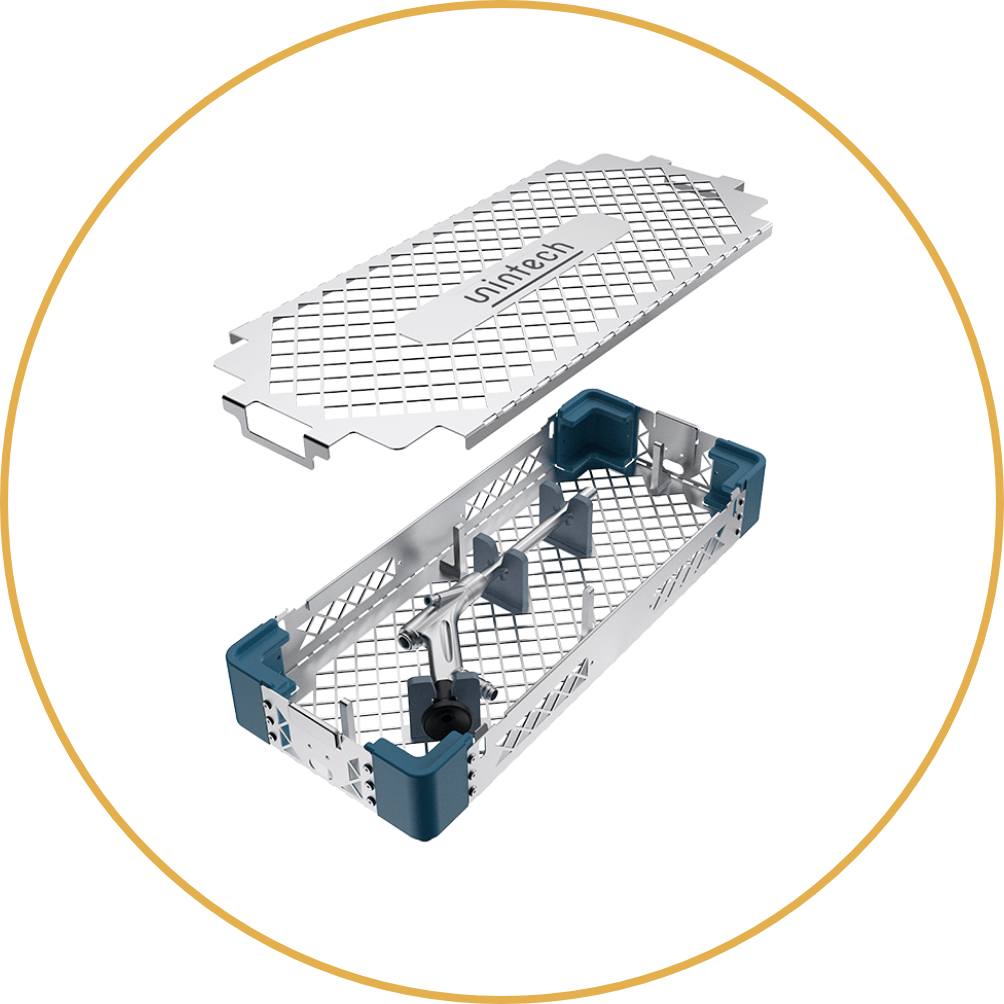
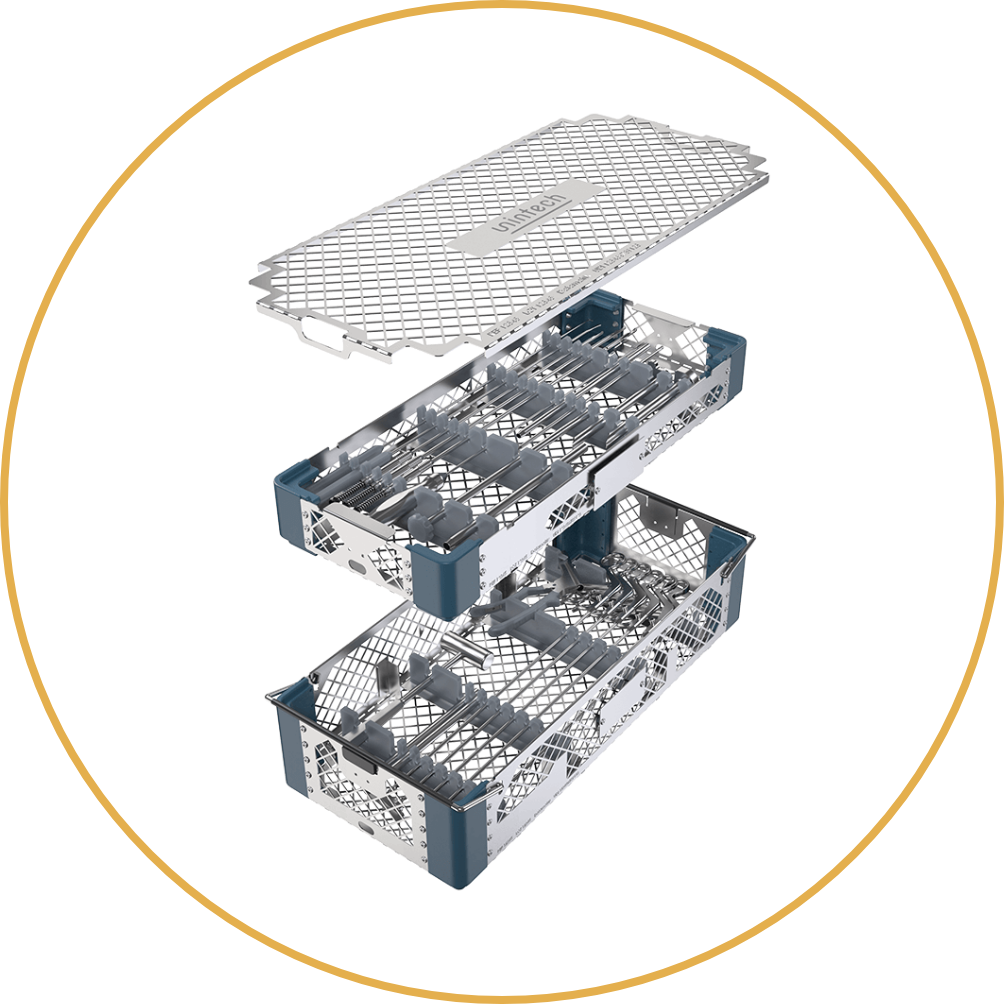
EndoSurg Standard System

transforaminal
  • Access: transforaminal and interlaminar
  • Indications: ventral and lateral pathologies, e.g. herniated discs, foraminal stenosis, facet joint cysts, spondylolistheses
  • Field of application: lumbar and thoracic

The EndoSurg Standard System is unintech‘s transforaminal system for full-endoscopic spinal decompression.

The system consists of instruments clearly arranged in a double tray and the endoscope safely stored in a tray, which also has space for the endoscopic light guide.

The set contains all instruments (approx. 22), that are required as standard for endoscopic surgery, e.g. dilators, working tubes, various forceps, chisels, nerve hook, elevator, etc. They are optimally adapted to the procedure and the endoscope in terms of length, diameter and design.

EndoSurg Standard Endoscope  
Working length 176 mm
Outer diameter shaft 6.6 mm
Diameter working channel 3.7 mm
Optic angle 30°
EndoSurg Endoscope

Principal steps of the procedure

Endoscopic pictures show transforaminal approach

Inserting the working tubes U and T
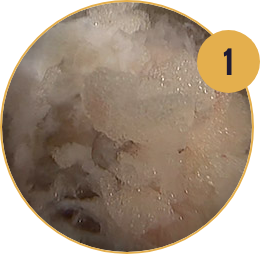
  1. Transforaminal access to the facet joint through serial Dilation
Endoscopic decompression with various instruments
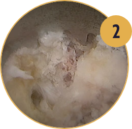
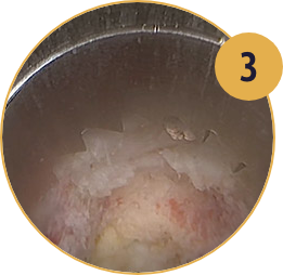
  1. Inserting the working tubes and identifying the intact and untreated bony structures. The outer working tube with teeth is fixed at the bone
Endoscopic view to intervertebral foramen
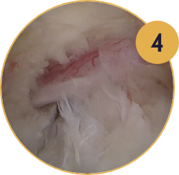
  1. Foraminotomy with forehead reamer under endoscopic visualization and monitoring
Endoscopic decompression with various instruments
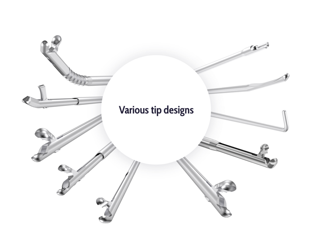
  1. Decompression of the nerves with various instruments
Endoscopic decompression with various instruments
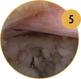
  1. End of the procedure - free nerves

Unique Instruments of the System

The transforaminal approach can be done with the unique Tube-in-Tube technique or traditonal in single tube technique.

  • Single tube technique: with the inner working tube
  • Tube-in-Tube technique: with both working tubes
  • Outer working tube with partial toothing
    Outer working tube with
    partial toothing
  • Working tube, oval window
    Working tube, oval window
    (inner working tube)

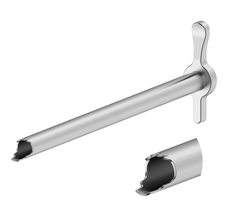
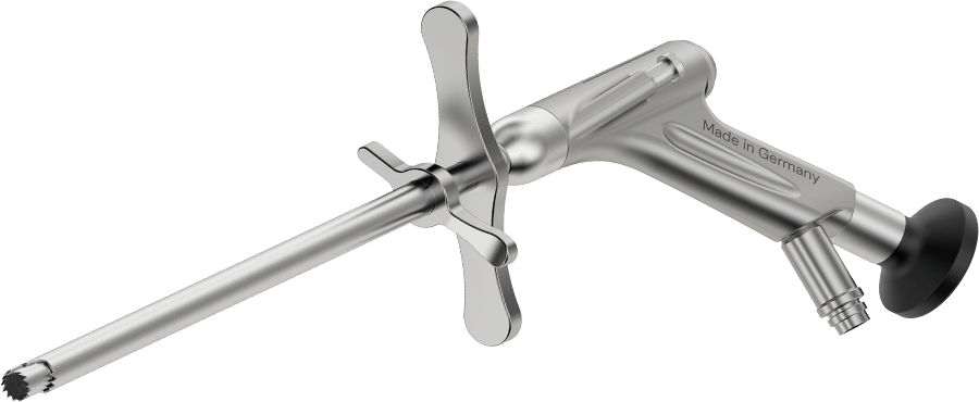
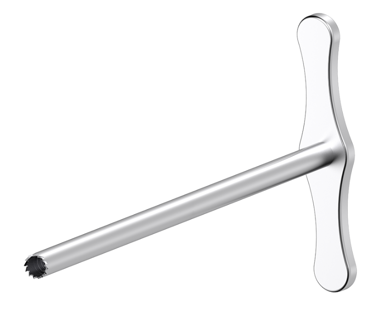
The outer working tube has teeth for fixation at the facet joint.
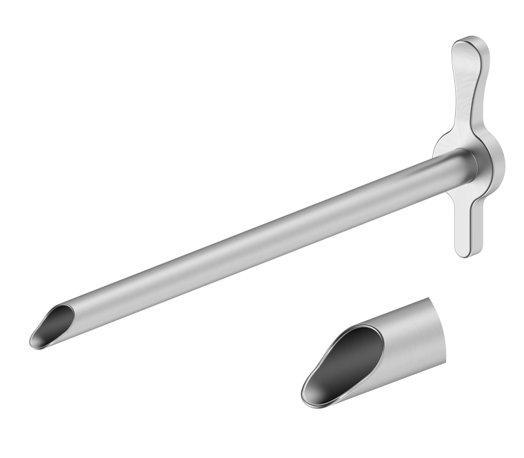
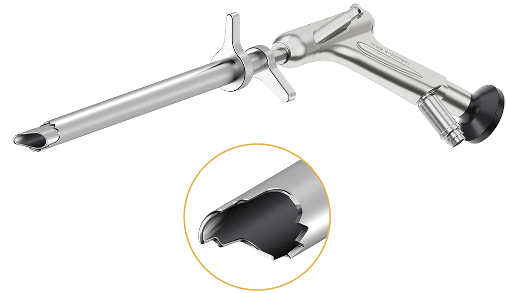
 The inner working tube can be used as retractor.

  • Forehead reamer
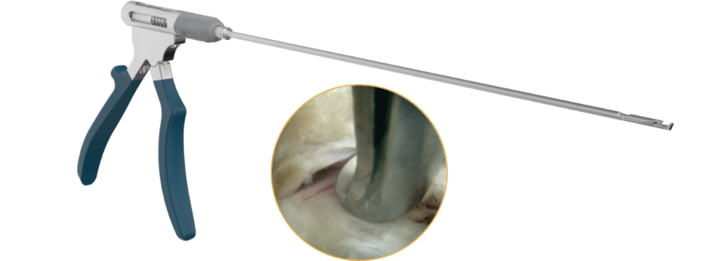
    Forehead reamer for endoscopic
    visualized foraminotomy
  • working tube endoscope
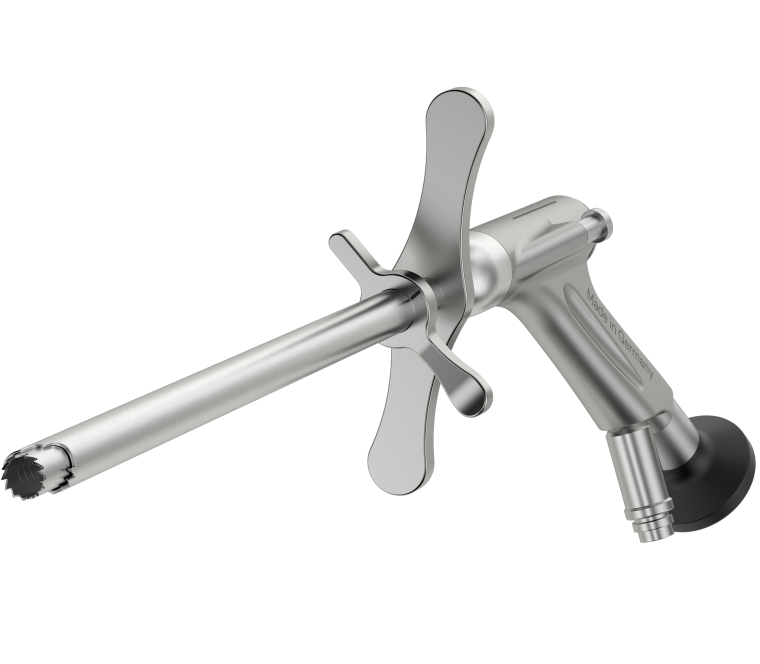
    Endoscope inserted in forehead
    reamer and outer working tube

Tube-in-Tube technique: Only with this technique endoscopic visualized and monitored foraminotomy is possible. The inner working tube is replaced by a forehead reamer

EndoSurg Standard Instruments for Decompression

  • Nerve hook and elevator for palpation
  • Nerve hook bone chisel and curette for boney treatment, e.g. osteophytes
  • Endoscopic Kerisson punches with rotatable shaft and various footprint
  • Forceps with different tips and sizes available, resilient and durable due to new and patented functional mechanism
Endoscopic Kerrison
Kerisson punch cutting Lig. flavum, transforaminal access
Scroll to Top